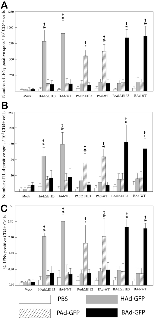
Figure 1. Cross-reactivity of positively selected CD4+ splenocytes from HAdΔE1E3-, PAdΔE1E3-, BAdΔE1E3-, HAd-WT-, PAd-WT-, BAd-WT-, or mock-inoculated mice.
Splenocytes were positively selected with anti-CD4 monoclonal antibody-coated magnetic beads and were stimulated with HAdΔE1E3-, PAdΔE1E3-, BAdΔE1E3-, or mock-infected cell lysate for 20 h. The number of cells expressing IFNγ (A) or IL-4 (B) was measured by enzyme-linked immunospot (ELISPOT) assay. (C) The percentage of CD4+ cells expressing IFNγ was measured by flow cytometry assay. Values are reported as the average ± standard deviation for five animals per group. *P < 0.005 versus values at mock stimulation within each treatment group. †P < 0.005 for homologous stimulation versus heterologous stimulation within each treatment group.

