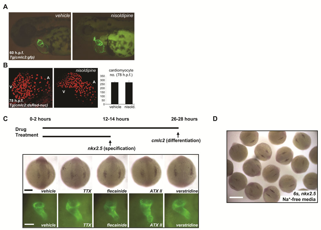
FIGURE 6. Voltage-gated sodium channels may regulate early cardiac development independent of membrane electrophysiology.
A. Rearing of embryos in 10µM solution of the L-type calcium blocker nisoldipine inhibited heart-beating at all stages examined through 78hpf but did not perturb early chamber formation or the initiation of looping. B. Analysis of vehicle-treated and nisoldipine-treated Tg(cmlc2:DsRed2-nuc) embryos indicated that L-type calcium channel-blockade did not affect embryonic cardiomyocyte number through 78hpf. Results are mean ± s.e.m for vehicle-treated (n = 2) and nisoldipine-treated (n = 3) embryos, respectively. C. Prolonged exposure of developing embryos to pharmacological modulators of sodium channel function failed to disrupt either cardiac specification or differentiation. In situ hybridization for nkx2.5 at the 6–8 somite stage (top) and fluorescent microscopy of the heart tubes of Tg(cmlc2:GFP) embryos at 26–28hpf (bottom), after the indicated treatments (see Table 2). TTX = tetrodotoxin, ATX II = anemone toxin II. Scale bars = 200µm (top) and 100µm (bottom). D. In situ hybridization for nkx2.5 at the 6 somite stage after incubation from the 1 cell stage in sodium-free media, to inhibit inward sodium current (INa). Early heart development proceeded normally. Heart tubes also form normally (not shown). Scale bar = 500µm.

