
Figure 9. Ion Channels in PAH.
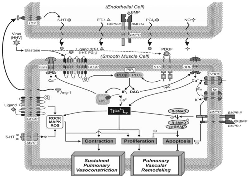
Schematic depiction of the cellular mechanisms linked with the vasoconstriction and remodeling in pulmonary endothelial (PAEC) and PASMC in PAH. Central themes of interest for the development of PAH include: 1) impaired ion channel expression and function in PASMC (Kv, VDCC, SOC, ROC), 2) increased cytosolic calcium ([Ca2+]cyt) in PASMC (mediated by ion channel function and receptor stimulation), 3) altered signaling via membrane receptors (GPCR, TIE-2, BMPR, RTK) and transporters (i.e., SERT) in endothelial cells and PASMC, 4) changes in redox status, 5) enhanced production of vasoconstrictor or mitogenic factors, and 6) viral signaling via GPCR and RTK. Paracrine interactions between PAEC and PASMC are noteworthy.
Abbreviations: Ang-1, angiopoietin-1; ET-1, endothelin-1; GPCR, G protein-coupled receptor; 5-HT, serotonin; MAPK, mitogen-activated protein kinase; NO, nitric oxide; PDGF, platelet-derived growth factor; PGI2, prostaglandin I2; ROC, receptor-operated Ca2+ channels; ROCK, Rho-associated kinase; ROS, reactive oxygen species; RTK, receptor tyrosine kinase; SERT, 5-HT transporter; SOC, store-operated Ca2+ channels; SR, sarcoplasmic reticulum; VDCC, voltage-dependent Ca2+ channels.
Figure and Legend Contributed by Drs. Carmelle Remillard and Jason Yuan, University of California, San Diego