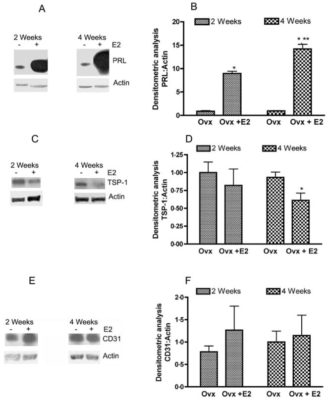
Figure 3.
Estradiol-induced changes in the levels of PRL, TSP-1, and CD31 proteins in the pituitary tissues. Pituitaries of F344 rats that were ovariectomized and treated with an empty implant (Ovx) or estradiol implant (Ovx + E2) for 2 and 4 weeks were used for measurement of PRL (A and B), TSP-1 (C and D), and CD31 (E and F) levels by western blot. Left panels show representative blots. Right panels show the mean ± S.E.M. ratios of band intensities of proteins (PRL, TSP-1, and CD31) and actin. Each bar represents mean ± S.E.M. of four different animals. *P<0.05 compared with the Ovx group; **P<0.05 compared with the Ovx + E2 treated at 2 weeks groups.

