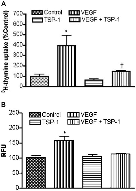
Figure 5.

Effect of TSP-1 on basal and VEGF-induced proliferation and migration of endothelial cells. (A) Cell proliferation assay. Endothelial cells grown in 96-well plate were treated with TSP-1 (5 μg/ml), VEGF (50 ng/ml), or VEGF and TSP-1 for 24 h. The cultures were treated with 0.5 μCi[methyl-3H]-thymidine per well for 8 h prior to harvesting. Data are presented as percentage of control. Each bar diagram represents mean ± S.E.M.; n=5–6. *P<0.01 compared with control group. †P<0.05 compared with VEGF-treated groups without TSP-1. (B) Cell migration assay. Equal numbers of serum-starved cells were plated on the migration tray with filter (8 μm pore size). To the lower chamber, TSP-1 (5 μg/ml) and VEGF (50 ng/ml) were added alone or together. Cell migration in each chamber was determined. The data are presented in relative fluorescent units (RFU). Each bar diagram represents mean ± S.E.M.; n=4. *P<0.01 compared with the rest of the groups.
