
Fig. 3.
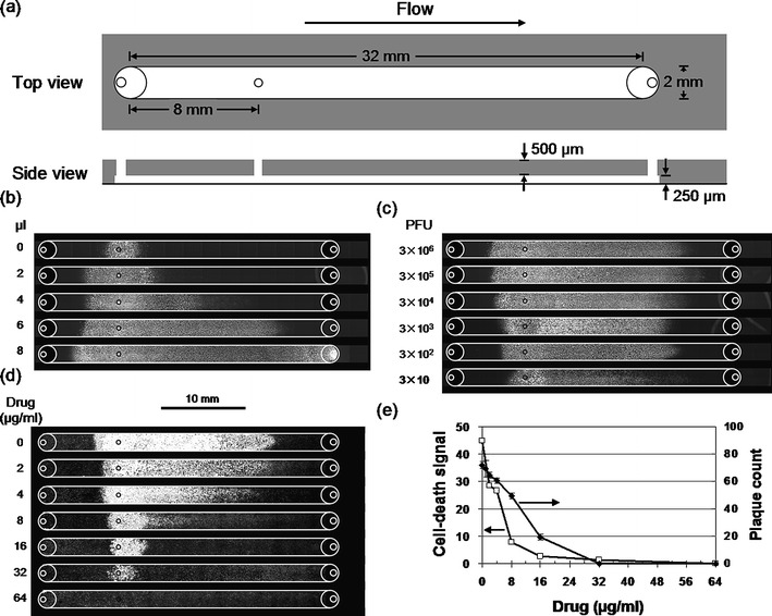
Fluid flow enhanced the spread of virus infections in microchannels. (a) Schematic of channel for virus infections in the presence of fluid flow. An inoculation port (diameter 0.5 mm) near the inlet port enabled localized initiation of infections. Volumes were 16 μl (channel) and 2 μl (each port). (b) Larger bolus volumes enhanced infection spread. Cell monolayers were locally inoculated with virus, incubated, subjected to bolus flows, further incubated, and GFP signals were detected. (c) Smaller inoculum sizes (PFU) reduced infection spread. Inocula containing 300 to 3 million PFU produced similar patterns of infection-mediated GFP expression for 6 μl bolus flows. However, an inoculum containing 30 PFU spread a shorter distance (p = 0.0005) and its GFP intensity was lower (p = 0.0008) than the others. (d) Higher drug levels inhibited virus infection spread. Cell monolayers were inoculated with 300 PFU, and both inocula and bolus flows contained the anti-viral drug 5-fluorouracil (5-FU). Regions of cell death (white) were visualized by staining with crystal violet. (e) Microfluidic infection assay was more sensitive than the plaque-reduction assay. Levels of drug treatments that were needed to achieve half-maximal infection (IC50) were 5 μg/ml (microfluidic infection assay) and 12 μg/ml (plaque-reduction assay)