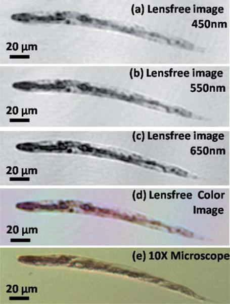
Fig. 5.
(a, b, and c) Digitally reconstructed holographic images of a stained C. elegans sample using Ponceau S red stain, captured with illumination wavelengths at 450 nm, 550 nm and 650 nm, respectively (FWHM ≈ 15 nm in each case). (d) Lensfree color image obtained by fusing the reconstructions at each wavelength. (e) Brightfield microscope image obtained with a 10× objective-lens for comparison purposes.

