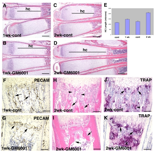
Fig. 9.
Inhibition of MMP activity inhibits residual endochondral ossification at the MMP-9−/− growth plate. (A–D) H&E-stained sections of metatarsals of 1-week-old MMP-9−/− mice treated for 1 week (A,B) or 2 weeks (C,D) with either vehicle or the MMP inhibitor GM6001, showing further lengthening of the HC (hc) zone in the GM6001-treated mice (B,D) compared with the vehicle-treated mice (A,C). (E) Bar graphs of the mean and standard deviation of the HC zone lengths of six control and GM6001-treated bones after 1 and 2 weeks of treatment; the differences between control and treated samples at each time point were statistically significant (P<0.01). (F,G) Immunostaining of tissue sections with PECAM-1 antibody showing metaphyseal vessels running parallel to hypertrophic chondrocyte columns in the vehicle-treated mice (F, arrows) and dilated vessels running perpendicular to the hypertrophic chondrocyte columns in the GM6001-treated mice (G, arrows). (H,I) H&E-stained sections showing primary (H, arrowheads) and secondary (H, arrows) trabeculae in the vehicle-treated mice, and a mass of bone instead of secondary trabeculae in the GM6001-treated mice (I, arrows). (J,K) TRAP staining of tissue sections showing the presence of TRAP+ cells at the cartilage-bone junction (arrows) and on trabecular bone surfaces (arrowheads) in both vehicle-treated (J) and GM6001-treated (K) mice. Bars, 400 μm (A–D); 100 μm (F–K).

