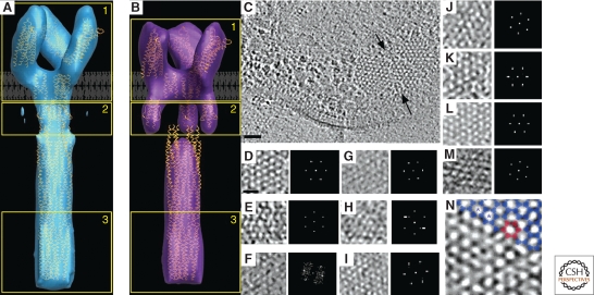
Figure 6.
Chemotaxis. (A) Compact and (B) Expanded averaged conformations of the TsrQEQE receptor. The structural coordinates corresponding to chemoreceptor models are loosely fit to a single trimer of receptor dimers. Adapted from (Khursigara et al. 2008b) with permission. © 2008 National Academy of Sciences, USA. (C) Hexagonal arrangement of receptors in different bacteria. Top view of a chemoreceptor array (between black arrows) in Thermotoga maritima. A subregion of the hexagonally ordered lattice and its corresponding power spectrum showing the ∼12 nm periodicity are enlarged in the inset. Scale bar 50 nm. (D–M) Top views of receptor arrays in other organisms. (D) T. maritima; (E) A. longum; (F) C. jejuni; (G) H. hepaticus; (H) M. magneticum; (I) H. neapolitanus; (J) R. sphaeroides; (K) E. coli; (L) V. cholerae; (M) T. primitia. Scale bar 25 nm, power spectra enlarged. (N) Trimer of dimers (blue) fit into the vertices of the hexagonal lattice in a chemoreceptor array. Six trimers of dimers (red) enclose one hexagon. The spacing from the center of one hexagon to another (blue asterisks) is 12 nm. Adapted from (Briegel et al. 2009) with permission. © 2009 National Academy of Sciences, USA.

