Figure 3.
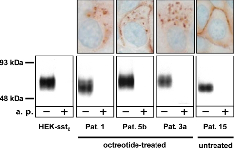
Confirmation by immunoblotting (bottom row) of the sst2 expression detected with sst2 immunohistochemistry (top row) in tumor samples of octreotide-treated patients (patients 1, 5b, and 3a) compared with controls consisting of sst2-expressing HEK293 cells (HEK-sst2) and a tumor sample from a patient not treated with octreotide (patient 15). In the immunoblot, a broad band centered around 70 kDa is stained by the R2-88 antiserum in all tested samples. This staining is completely abolished by preadsorption of R2-88 with the antigen peptide (a.p.). The variation in apparent molecular mass of the sst2 receptor in the analyzed samples reflects the differences of glycosylation of the receptor. The positive sst2 staining detected with the R2-88 antiserum in the immunohistochemistry samples of patients 1, 5b, and 3a as well as the membranous staining seen in the sample of patient 15 together with the presence of a sst2 receptor band in all samples detected by R2-88 immunoblotting strongly suggests that the granular cytoplasmic structures seen by immunohistochemistry indeed correspond to the internalized sst2 receptor. Pat., Patient.

