Fig.
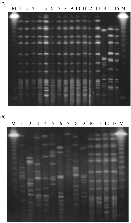
Macrorestriction analysis of chromosomal DNA derived from Legionella pneumophila sg 1 and sg 5 digested with SfiI and separated by pulsed-field gel electrophoresis. (a) A comparison of the clinical isolates with the strains isolated from water samples and swabs at different sites of the ship. Lanes 1 and 2, sg 5 isolates from patient (case 1) sputum; lane 3, sg 5 isolate from men‘s spa water; lanes 4–8, sg 5 isolates from natural stones in men’s spa filter; lanes 9–12, sg 5 isolates from natural stones in women’s spa filter; lane 13, sg 5 isolate from a strainer of women’s spa; lane 14, sg 5 isolate from a whirlpool spa; lane 15, sg 1 isolate from a swab at a strainer for women’s spa; lane 16, sg 1 isolate from natural stones in women’s spa filter. (b) L. pneumophila sg 5 strains. Lane 1, NIIB 412, Osaka LG02-11 from a spa bath; lane 2, ATCC 33216 Dallas 1E from a cooling tower; lane 3, NIIB 98 (EY 3420), a clinical isolate in Osaka [22]; lane 4, NIIB104 (EY 3427), a clinical isolate in Kurashiki; lane 5, ATCC 33737 U8W from shower head water; lane 6, NIIB 288 Ishioka 1-2-4 from a spa bath [18]; lane 7, NIIB 330 (ThaiNIH 7811) from a cooling tower; lane 8, NIIB 361 (ThaiNIH 10723) from a cooling tower; lane 9, corresponding to lane 14 of panel (a); lane 10, corresponding to lane 1 of panel (a); lane 11, corresponding to lane 4 of panel (a); lane 12, corresponding to lane 5 of panel (a); lane 13, corresponding to lane 6 of panel (a). Ms are DNA size markers, lambda ladders (Bio-Rad, Richmond, CA, USA), as indicated on the right and left sides of each electrophoregram.

