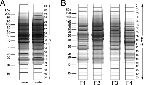
Figure 3.
SDS-PAGE separation of melanoma 1205Lu cell lysate and MicroSol fractions for proteome analysis. Samples were electrophoresed until the tracking dye migrated 4 cm; gels were stained with Colloidal Coomassie and individual lanes were cut into 20 equal-sized slices, as shown. (A) For the 2-D method, the unfractionated lysate of the 1205Lu cells was separated in two lanes (60 μg/lane). The supernatants from corresponding slices in the two lanes were combined after trypsin digestion. (B) MicroSol IEF fractions derived from 120 μg of cell lysate were separated for the 3-D method.

