Figure 1.
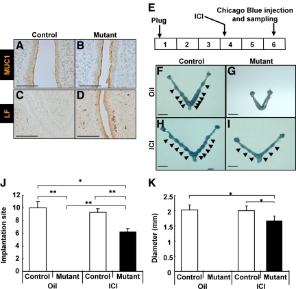
ICI-rescued implantation defect of PRCre/+;COUP-TFIIflox/flox mutant mice. A–D, ERα activity, as examined by the expression of MUC1 and LF, was elevated in the epithelium of COUP-TFII mutant mice. A and B, Expression of MUC1 in the uterine epithelium was elevated in mutant mice at 30 h after eP treatment. C and D, Expression of lactoferrin in the uterine epithelium was elevated in mutant mice at 30 h after eP treatment (scale bar, 100 μm). E, Experimental scheme of rescue experiment for COUP-TFII mutant implantation defect by ICI injection. F–I, Implantation occurred in PRCre/+;COUP-TFIIflox/flox mutant mice treated with ICI. Implantation sites were visualized as blue spots (arrowhead) by iv injection of Chicago blue dye at d 6. F, Control mice treated with oil. G, Mutant mice treated with oil. H, Control mice treated with ICI. I, Mutant mice treated with ICI (scale bar, 5 mm). J, Average number of implantation sites of rescue experiment. ICI rescued implantation defect of mutant mice although the number is slightly reduced. All mutant mice were rescued after ICI treatment (control mice with oil, n = 3; mutant mice with oil, n = 3; control mice with ICI, n =3; mutant mice with ICI, n = 4). K, Implantation sites of ICI-treated mutant mice were only a bit smaller than control mice treated with either oil or ICI. White bars, control; black bars, mutant. *, P < 0.01; **, P < 0.005.

