Figure 3.
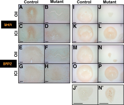
Decidual cells in ICI-treated mutant mice expressed decidual cell markers, WNTt4 and BMP2. A–D, Decidual cells surrounding implantation sites expressed decidual cell marker, WNT4 at d 6. A, Control mice treated with oil. B, Mutant mice treated with oil. C, Control mice treated with ICI. D, Mutant mice treated with ICI (longitudinal section: scale bar, 200 μm). E–H, Decidual cells surrounding implantation sites expressed decidual cell marker, BMP2 at d 6. E, Control mice treated with oil. F, Mutant mice treated with oil. G, Control mice treated with ICI. H, Mutant mice treated with ICI (longitudinal section: scale bar, 200 μm). I–L, Decidual cells in artificial decidualized uterine horns expressed decidual cell marker, Wnt4. I, Control mice treated with oil. J and J′, Mutant mice treated with oil. K, Control mice treated with ICI. L, Mutant mice treated with ICI (scale bar, 635 μm). M–P, Decidual cells in artificial decidualized uterine horns expressed decidual cell marker, BMP2. M, Control mice treated with oil. N and N′, Mutant mice treated with oil. O, Control mice treated with ICI. P, Mutant mice treated with ICI (scale bar, 635 μm).

