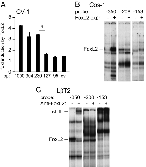
Figure 6.
FoxL2-binding sites in the −230/−127 region of the mouse FSHβ promoter. A, CV-1 cells were transfected with different lengths of the mouse FSHβ promoter indicated under the corresponding bar, with the FoxL2 expression vector or empty vector (ev) control. Fold induction by FoxL2 overexpression over empty vector control for each truncation are represented. *, Significant decrease in fold induction with P < 0.05. B, Probes encompassing the FoxL2-binding elements were incubated with nuclear extracts from Cos-1 cells transfected with FoxL2 (FoxL2 expr.) or ev control (−). C, Probes encompassing putative FoxL2-binding elements were incubated with nuclear extracts from LβT2 with or without inclusion of the FoxL2 antibody (Anti-FoxL2).

