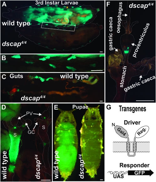
Figure 4.—
(A) Ventral views of dSREBP activity in wild-type (upper) and dscap4 (lower) third instar larvae visualized using the GAL4-dSREBP/UAS-GFP reporter system. Fluorescence is readily detected in dscap4 oenocytes, and to a lesser extent in regions of the anterior midgut. Larvae are wild type or homozygous for dscap4 on the second chromosome and homozygous for P{GAL4-dSREBPg}, P{UAS-GFP} on the third chromosome. Dashed lines denote extent of larval bodies. Exposure = 10 sec. (B) Higher magnification view of the region indicated by the white box in A showing wild-type and dscap4 oenocytes. Exposure = 4 sec. (C) Midguts showing reduced fluorescence in dscap4 larvae compared to wild type. Exposure = 8 sec. (D) Anterior midguts from wild-type and dscap4 larvae showing greatly reduced dSREBP activity in dscap4 larvae. GC, gastric caeca; PV, proventriculus; S, stomach; exposure = 4 sec. (E) Wild-type and dscap4pupae (stage P5) (Bainbridge and Bownes 1981). Exposure = 5 sec. (F) Anterior midgut from a dscap4 larva exhibiting fluorescence only at the posterior end of the proventriculus. Exposure = 3 sec. (G) Schematic of the GAL4-dSREBP; UAS-GFP reporter system. Scale bars, 0.2 mm. The variability of dSREBP activity in the midgut of dscap4 larvae is evident among the different individuals shown in C, D, and F (see also Figure 6A).

