
Figure 1.
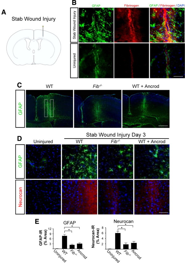
Fibrinogen depletion decreases astrogliosis and neurocan expression. A, SWI, a model of cortical trauma. B, Three days after SWI, immunolabeling for fibrin (red) and GFAP (green) revealed perivascular fibrin colocalizing with reactive astrocytes in brain coronal sections of mice (top). Uninjured mice show no fibrinogen deposition in the brain (bottom). C, Low-magnification images of Fib −/− and ancrod-depleted WT mice show reduced astrogliosis demonstrated by immunolabeling of GFAP astrocytes (green) 3 d after SWI. Box indicates area of quantification. D, Fib −/− and ancrod-treated WT mice show reduced astrogliosis and neurocan expression 3 d after SWI, shown by immunolabeling of astrocytes for GFAP (green) and neurocan (red). Uninjured WT mice served as controls. E, Quantification revealed lower levels of GFAP and neurocan in Fib −/− and ancrod-treated mice than in WT mice (n = 5 per group) after SWI. Values are mean ± SEM. *p < 0.001. Scale bars: B, 75 μm; C, 500 μm; D, 60 μm.