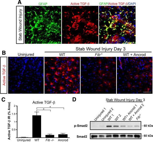
Figure 5.
Fibrinogen is necessary for active TGF-β formation and signaling in the CNS. A, Immunolabeling for GFAP (green) and active TGF-β (red) revealed astrocyte-specific expression of active TGF-β after SWI (yellow). B, Decreased levels of active TGF-β immunolabeling (red) in Fib −/− and ancrod-treated mice 3 d after SWI. Uninjured WT brain served as a negative control. C, Lower levels of active TGF-β after SWI in Fib −/− and ancrod-treated mice than in WT controls (n = 5 per group). D, Brain lysates of Fib −/− and ancrod-treated WT mice show reduced Smad2 phosphorylation 3 d after SWI. Brain lysates of uninjured WT mice do not show P-Smad2 activation. Lysates from two mice per experimental treatment are shown. Values are mean ± SEM. *p < 0.001. Scale bar, 60 μm.

