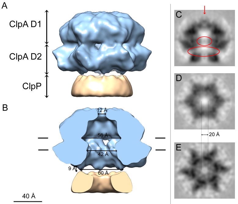
Figure 2.
Cryo-EM 3D reconstruction of ClpA. (A) Surface rendering of the side-view and (B) cut away view exposing the interior. The ClpA hexamer is in blue and a portion of ClpP (~25%) is in ochre: the latter appears almost perfectly cylindrically symmetric because 6-fold symmetrization has been applied to its 7-fold-symmetric structure. The D1 and D2 tiers of ClpA are labeled. Interior dimensions at various locations are marked. In A and B, a small diffuse density plugging the apical entrance to the axial channel (red arrow in panel C) has been removed for clarity. (C–E) Grayscale sections; (C) Central longitudinal section for the plane contoured in B. D and E are transverse sections, looking towards ClpP. D is at the level of the “56 Å” channel in B, and F is at the “42 Å” mark. The upper red oval in C marks low but significant axial densities visualized within the ClpA channel that represent mobile elements. Although the surface rendering in B depicts cavity diameters of ~ 56 Å in D1 and ~ 42 Å in D2, these measures are reduced to ~ 20 Å for the completely open channel in the corresponding grayscale sections (D and E respectively). The bottom red oval encloses densities bridging between the ClpA and ClpP rings that have been diluted by applying 6-fold symmetry to the ClpP heptamer in the reconstruction.

