
Fig. 2.
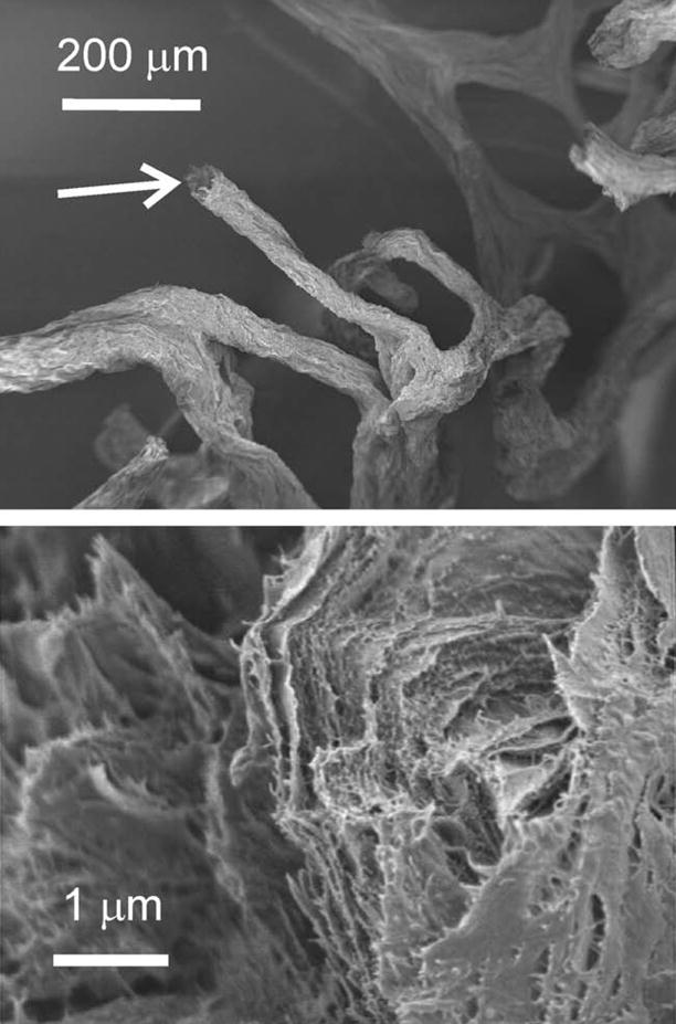
SEM images showing the micro- and nano-organization of a selected chitin fiber from the I. basta skeleton after step 3. The image at the bottom is taken from the disrupted fiber shown in the top image (white arrow).
Official websites use .gov
A
.gov website belongs to an official
government organization in the United States.
Secure .gov websites use HTTPS
A lock (
) or https:// means you've safely
connected to the .gov website. Share sensitive
information only on official, secure websites.
SEM images showing the micro- and nano-organization of a selected chitin fiber from the I. basta skeleton after step 3. The image at the bottom is taken from the disrupted fiber shown in the top image (white arrow).