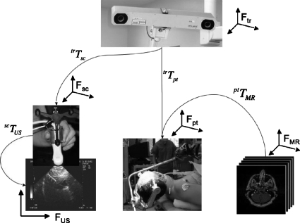
Figure 1.
Overall view of the various components of image guidance involving freehand tracked ultrasound. At the top is the 3D tracker. The lower left image shows the ultrasound scanhead, its rigidly coupled tracker, and the image it produces. The lower middle image shows a patient and the tracker coupled to the head clamp in the OR. The lower right images show the preoperative MRI imaging study. The arrows indicate how the various frames of reference are related and the respective transformation matrices that are involved. The corresponding frames of reference are also shown (f) near each image.

