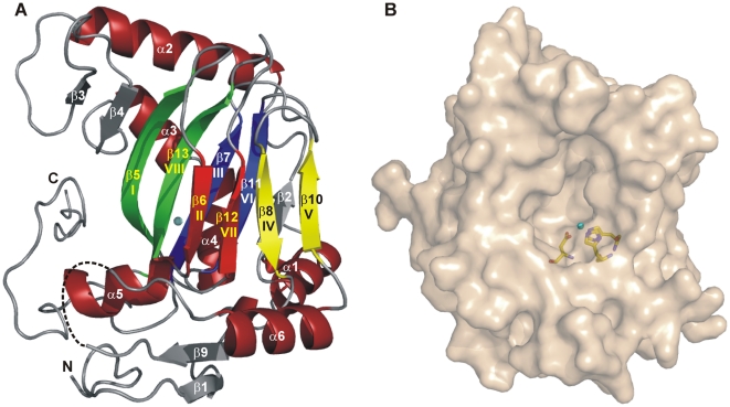
Figure 2. Ribbon and surface representation of the EctD ectoine hydroxylase.
(A) The successive segments of the double-stranded β-helix (DSBH) are coloured according to the scheme of Branden and Tooze [75]. The Fe3+ ion bound by EctD is shown as a blue sphere. A dashed line indicates a disordered loop region connecting the DSBH β-strands IV and V. (B) The surface of the EctD protein is represented and the side-chains of the iron-coordinating residues His-146, Asp-148 and Asp-248 are shown as sticks. The iron ion bound by the crystallized EctD protein is shown as a blue sphere.

