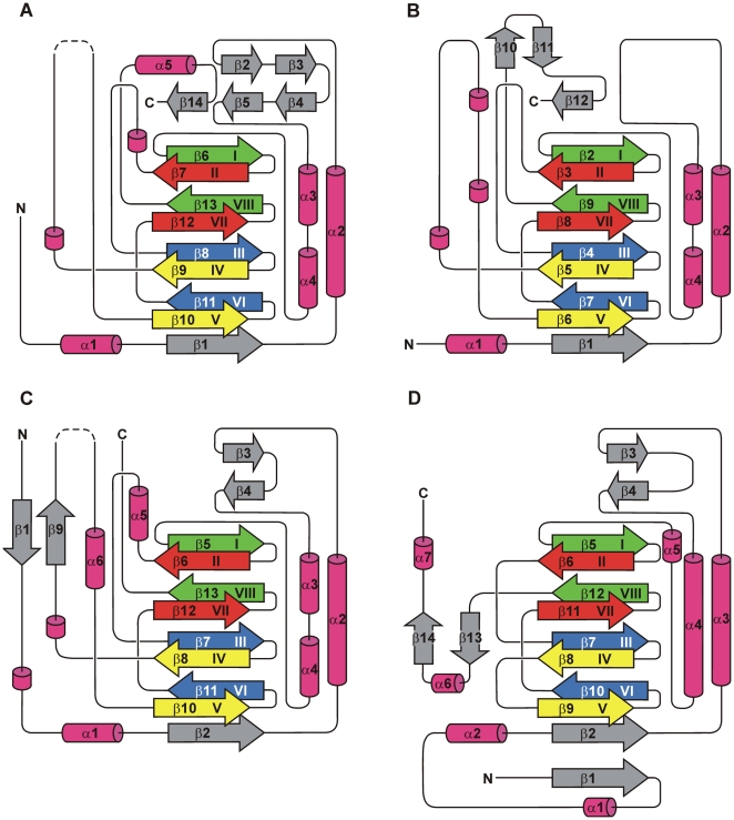
Figure 3. Topology diagrams of PhyH-family enzymes PhyH, SyrB2 and EctD and of the DAOCS enzyme.
(A) PhyH (PDB code: 2A1X), (B) SyrB2 (PDB code: 2FCU) and (C) EctD (PDB code: 3EMR). (D) Topology diagram of DAOCS (PDB code: 1DCS), a representative of the family of the IPNS/DAOCS-like enzymes. Arrows represent β-strands, while α-helices are shown as cylinders. 310-helices are also shown as cylinders, but not numbered. The β-strands forming the DSBH-motif are coloured according to the scheme of Branden and Tooze [75]. In the diagrams of EctD and PhyH, the disordered putative “lid” region within the segment connecting DSBH β-strands IV and V is shown by dashed lines. For the sake of clarity and comparability, DSBH β-strand II is included in the topology diagram of PhyH, although this strand is largely disordered and invisible in the corresponding crystal structure [40].

