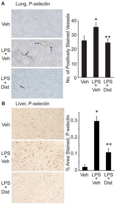
Figure 4. Distamycin A decreases lung and liver P-selectin tissue staining.
A. Representative sections (200× magnification) of lung tissue harvested from C57BL/6 mice 24 h after treatment with Vehicle (Veh), LPS+Veh, or LPS+Dist A (Dist). Tissue was fixed, processed, and stained with a P-selectin antibody. Number of positively stained vessels was counted in sections from at least 5 mice in each treatment group. (Arrows demonstrate examples of positively stained vessels; *p<0.05 compared with Veh; **p<0.05 compared with LPS/Veh). B. Representative sections (200× magnification) of liver tissue harvested from mice 4 h after treatment with Vehicle (Veh), LPS+Vehicle (Veh) or LPS+Dist A (Dist). Tissue was fixed, processed, and stained with a P-selectin antibody. Using NIH Image Software, 5 fields (at 200× magnification) per liver section from mice from each treatment group were quantified, with brown pixels counted as positive staining. Results were expressed as % positively stained area per 200× field. (*p<0.05 compared with Veh; **p<0.05 compared with LPS/Veh).

