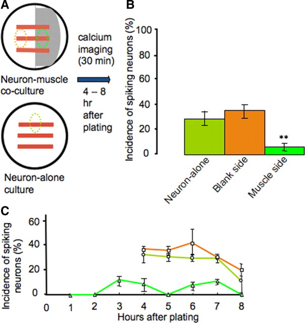
Figure 6.
Muscle cells suppress early neuronal spontaneous calcium spike activity. A, Diagram showing areas of interest in which neurons were imaged. B, Neurons grown on muscle cells exhibit a significantly lower incidence of spiking at 4–8 h in vitro (n = 10 cultures, >50 neurons per condition; **p < 0.01). C, Neurons grown on muscle cells exhibit reduced incidence of spiking during each hour after plating (n > 10 cultures, >50 neurons per hour). A higher percentage of neurons exhibit calcium spikes from 4 to 8 h on the blank side of cocultures and in neuron-alone cultures. Imaging at earlier times was not performed with the two control groups due to lack of morphological distinction between neurons before axon outgrowth and the presence of other cell types. Error bars indicate SEM. The Kruskal–Wallis test and Conover post hoc test were used to determine statistical significance.

