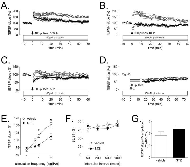
Figure 1. Diabetes impairs LTP at medial perforant path synapses in the dentate gyrus, and alters the threshold for induction of LTD.
(A), In agreement with previous reports (Kamal et al., 1999, Stranahan et al., 2008), diabetic rats showed reduced LTP at medial perforant path synapses on dentate granule neurons following a single, 1-second train delivered at 100 Hz. (B), Following a train delivered at 10 Hz, non-diabetic rats exhibit strong post-tetanic potentiation, with a gradual return to within 10–15% of baseline. In contrast, diabetic animals showed post-tetanic depression, followed by a return to baseline. (C), In response to a 5 Hz train, non-diabetic rats exhibit a slight posttetanic potentiation, followed by a return to baseline, while diabetic rats exhibit posttetanic depression and a return to baseline. (D), There was no difference in the magnitude of LTD following 900 pulses delivered at 1 Hz. (E), Summary graph showing the fEPSP slope as a percentage of pre-tetanus baseline, plotted against the log of the stimulation frequency. Asterisk (*) indicates significance at p<0.05 following ANOVA with Tukey’s post hoc. (E), Diabetes did not alter the paired-pulse depression that is characteristic of the medial perforant pathway (Colino and Malenka, 1993). (F), The ratio of the presynaptic fiber volley and the postsynaptic fEPSP was not influenced by diabetes, suggesting that there was no change in the input-output relationship.

