Fig. 3.
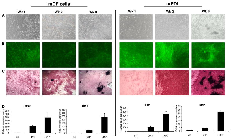
In vitro analysis of the osteogenic potential of primary dental follicle and periodontal ligament (PDL)-derived cells. Murine dental follicle (mDF) cells (left panel) derived from 3–5-d-old mice, and murine PDL (mPDL) cells (right panel) from 4–6-wk-old α-smooth muscle actin–green fluorescent protein (αSMA–GFP) transgenic mice were imaged using phase-contrast microscopy (A). The GFP expression of both mDF and mPDL cells was monitored at different time points and the corresponding stage of differentiation: week 1 (confluent cell stage), week 2 (multilayer formation stage) and week 3 (mineralized nodule stage) (B). The cells were also analyzed for alkaline phosphatase (ALP) activity at these three time points. ALP activity was detected at week 1 and was maximal at week 2 (C). von Kossa staining revealed the presence of mineralized nodules at week 3. The mDF and mPDL cells were analyzed for the expression of bone sialoprotein (BSP) and dentin matrix protein (DMP) by real-time PCR. Maximum expression of these genes was observed after 3 wk in culture along with an increase in mineralization (D). Images were taken under 10× magnification (bar = 200 μm).

