
FIGURE 3.
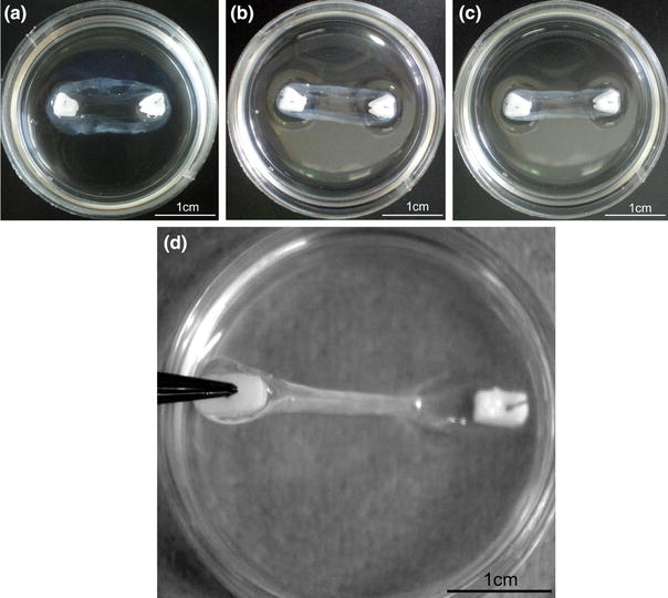
Formation and testing of brushite cement anchor/tissue interface. Tissue-engineered ligament formation with cell-seeded fibrin gel and brushite cement anchors (a) 3 days, (b) 5 days, (c) 7 days after plating as the fibrin gel contracts around the two cement anchor points. (d) Testing cement anchor/fibrin attachment. Cement anchor was lifted off the sylgard layer by the embedded pin. Attachment was scored as attached (as shown) or not attached if the soft tissue detached from the cement anchor