
Fig. 3.
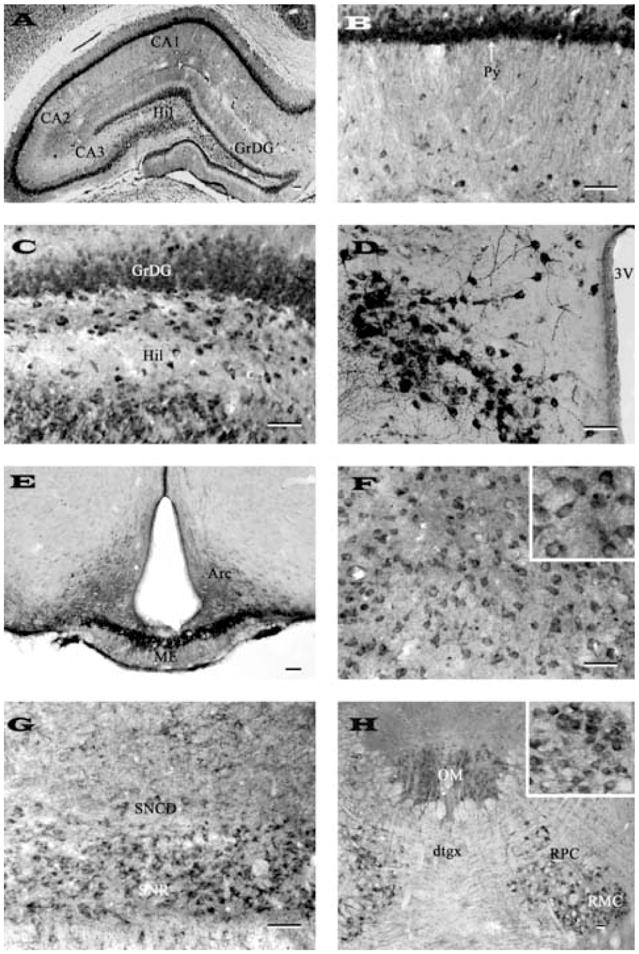
Photomicrographs of transverse sections of adult rat brain showing the distribution of nicastrin immunoreactive neurons and fibers in the hippocampus (A), CA1 pyramidal cell layer of the Ammon’s horn (B), granule cell layer and hilus of the dentate gyrus (C), paraventricular nucleus of the hypothalamus (D), arcuate nucleus/median eminence (E), medial thalamic nucleus (F), substantia nigra (G) and in oculomotor and red nuclei (H). Note intense labeling in neurons of the pyramidal cell layer, hilus and paraventricular nucleus and fibers in the median eminence. Inset in (F) and (G) show neuronal labeling at higher magnification. Representative photomicrographs of medial thalamic nucleus (F) and substantia nigra (G) were obtained with N-19 antiserum, whereas others (A, B, C, D, E, H) were acquired following labeling with SP718 antiserum. Scale bar = 50 μm. Hil, hilus; GrDG, granular cell layer of the dentate gyrus; Py, pyramidal cell layer; 3V, third ventricle; Arc, arcuate nucleus; ME, median eminence; SNCD, substantia nigra compact part; SNR, substantia nigra reticular part; OM, oculomotor nucleus; dtgx, dorsal tegmental decussation; RPC, red nucleus pervicellular part; RMC, red nucleus magnocellular part.