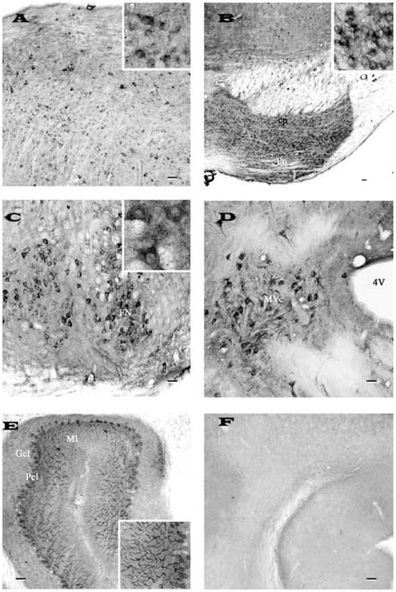
Fig. 4.

Photomicrographs of transverse sections of adult rat brain showing the distribution of nicastrin immunoreactivity in the inferior colliculi (A), pontine nucleus (B), facial nucleus (C), vestibular nucleus (D) of the brainstem and in the cerebellum (E). Note intense labeling of the brainstem neurons and the Purkinje cells of the cerebellum. F, represents a cerebellar section processed following preabsorption of the antibody with 10 μM purified rat nicastrin. Inset in (A), (B), (C) and (E) show neuronal labeling at higher magnification. All photomicrographs were acquired following labeling with SP718 antiserum. Scale bar = 50 μm. cp, cerebral peduncle; Pn, pontine nuclei; FN, facial nucleus; Mve, medial vestibular nucleus; Gcl, granular cell layer; Pcl, Purkinje cell layer; Ml, molecular layer.
