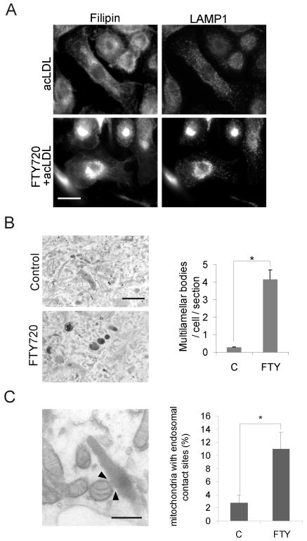
Figure 4. Late endosomal organelles are affected by FTY720.
A. Macrophages were treated ± 1 μmol/L FTY720 for 48 h and 50 μg/mL acLDL for the last 24 h of incubation. Cells were fixed and stained with filipin and anti-LAMP-1 antibodies. B. Electron microscopic images of cells treated ± 1 μmol/L FTY720 for 24 h, followed by a 2-h pulse with 50 μg/mL DiI-acLDL and a 4-h chase. The number of multilamellar bodies was quantified from 44 control cells and 48 FTY720 treated cells. Scale bar, 500 nm. C. Macrophages were treated for 24 h ± 1 μmol/L FTY720 prior to fixing and processing for electron microscopy. The percentage of mitochondria with endosomal contacts (example indicated by arrowheads) was quantified from 21 and 19 randomly chosen visual fields from control and FTY720 treated cells, respectively. Scale bar, 250 nm.

