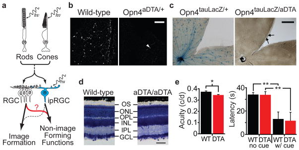
Figure 1. Elimination of ipRGCs in mouse retina.
a, Model describing how rod/cone signalling through conventional RGCs or ipRGCs contribute to NIF functions. Role of ipRGCs in image formation is speculative (dotted line). b, Melanopsin antibody staining in retinas of 18 month old wild-type (n=6) and Opn4aDTA/+ (n=12) mice. White arrowhead indicates a surviving ipRGC. Scale bar, 200μm. c, X-gal staining from Opn4tau-LacZ/+ (n=6) and Opn4aDTA/tau-LacZ (n=8). The surviving cells are weakly stained (black arrows). Scale bar, 500μm. d, Cross sections of Giemsa stained retinas from 18 month old Opn4aDTA/aDTA (aDTA/aDTA; n=3) and wild-type mice (n=3). The morphology of retinas is indistinguishable between genotypes. GCL, ganglion cell layer; IPL, inner plexiform layer; INL, inner nuclear layer; OPL, outer plexiform layer; ONL, outer nuclear layer; OS, outer segment. Scale bar, 50 μm. e, Left, the acuity of Opn4aDTA/aDTA mice (DTA; n=11; red bar) was slightly decreased compared to wild types (WT; n=9; black bar). Right, the latency to locate a marked platform (w/cue) in a water maze was similar between Opn4aDTA/aDTA (DTA; n=14; red bar) and wild types (WT; n=12; black bar). This latency significantly differed for unmarked platform (no cue) tests. All statistical comparisons utilized Student’s t test (*, p<0.05; **, p<0.01); error bars ± s.e.m.

