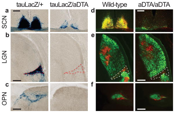
Figure 2. The ipRGC fibres in the brain decrease in aDTA mice.
a, b, and c, X-gal staining in Opn4tau-LacZ/aDTA (tauLacZ/aDTA; n=2) show that ipRGC innervation of the SCN, IGL and OPN is decreased. d, e, and f, Ocular cholera toxin injections (left eye, green; right, red) of Opn4aDTA/aDTA (aDTA/aDTA; n=11) and wild types (n=6). a and d, SCN innervation is sparse in aDTA mice. b and e, The dorsal lateral geniculate nucleus (LGN) is innervated similarly both in aDTA and wild-type animals, while few fibres remain in the IGL of mutant mice (outlined region). c and f, The OPN shell is innervated by ipRGCs and the core is targeted by other RGCs15. f, Fibres in the OPN core are retained in Opn4aDTA/aDTA mice. c, Fibres in the shell region are eliminated in Opn4tau-LacZ/aDTA animals. Scale bars, 200μm.

