Fig. 2.
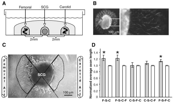
Directed migration in neurovascular co-cultures. (A) Schematic representing three-dimensional in-vitro co-culture. Whole SCG explants from postnatal day 2 mice were co-cultured in the presence of adult femoral and carotid artery segments. Tissues were embedded in type I collagen gel 2 mm apart and cultured for 16 h. (B) Tyrosine hydroxylase immunolabeling in whole SCG explants. Left, low magnification; right (boxed inset), high magnification. Scale bar=100 μm. (C) Representative image of SCG co-cultured with femoral and carotid artery segments. Femoral and carotid artery segments were placed 2 mm from SCG explants. To quantify outgrowth, the area occupied by the SCG and its neurites was divided into quadrants (solid lines). Average axon length was measured for 25 axons in the femoral and carotid directed quadrants (outlined by dashed lines). Carotid directed average axon length was normalized to 1 and femoral/carotid ratios were calculated. Scale bar=100 μm. (D) Directed neurite outgrowth of sympathetic axons from whole SCG explants. Neurites of equal average length grew radially from SCG alone controls (data not shown). In femoral/carotid co-cultures (first pair of bars) (FSC) the average axon length was significantly increased towards the femoral compared to the carotid artery. (n=14; *=p<0.05; vertical bars represent standard error.) To test the effect of vessel rearrangements on directed neurite outgrowth, SCG explants (S) were cultured in the presence of various arrangements of vessels. When femoral segments (F) were placed on the same side as carotid segments (C), either proximal (second pair of bars) (FSCF) or distal to the SCG (S) (third pair of bars) (CSFC) outgrowth towards the femoral segment was blunted, with the placement of carotid segments disrupting the typical increased outgrowth observed towards femoral segments. (n=14; & =p<0.05; vertical bars represent standard error.) When femoral segments (F) were placed on the same side as carotid segments (C), either distal (fourth pair of bars) (CSCF) or proximal to the SCG (S) (fifth pair of bars) (FSFC) outgrowth towards the carotid segment was unchanged.

