Figure 2.
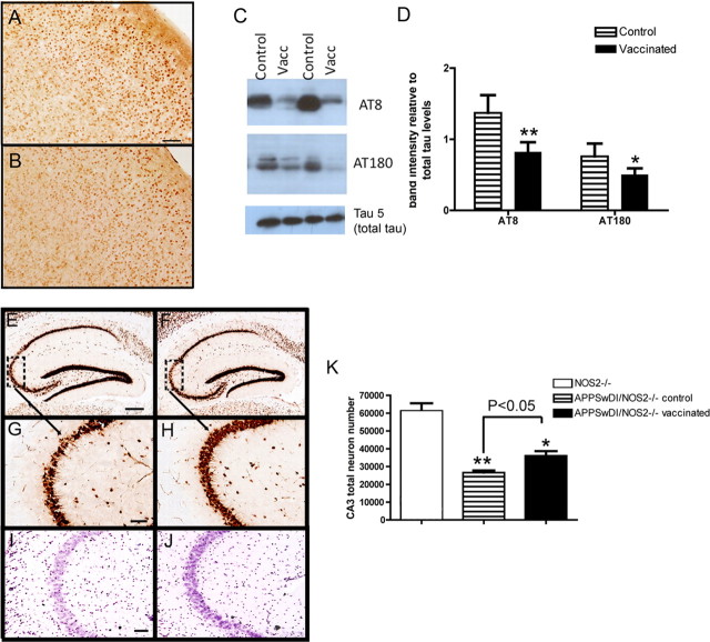
Tau pathology and neuron loss are reduced in APPSwDI/NOS2−/− mice receiving Aβ vaccination. A and B show AT8 immunohistochemistry in the cerebral cortex of APPSwDI/NOS2−/− mice receiving either control vaccination (A) or Aβ vaccination (B). Magnification, 200×; scale bar, 25 μm. C shows representative Western blots for AT8, AT180, and tau 5 from APPSwDI/NOS2−/− mice receiving control (control) or Aβ vaccination (Vacc). D shows mean densitometry data of AT8 and AT180 Western blots, normalized to total tau levels. N = 7 mice receiving control vaccination and N = 8 receiving Aβ vaccination. E–H show NeuN immunohistochemistry in the hippocampus (E and F, magnification, 40×; scale bar, 120 μm) and CA3 (G and H, magnification, 200×; scale bar, 25 μm) of APPSwDI/NOS2−/− mice receiving either control (E, G) or Aβ (F, H) vaccination. Dashed boxes in E and F indicate the region shown at higher magnification in G and H. I and J show cresyl violet staining of the CA3 of APPSwDI/NOS2−/− mice receiving either control (I) or Aβ (J) vaccination. Magnification, 200×; scale bar, 25 μm. K shows stereological counts of the CA3 region of NOS2−/−, APPSwDI/NOS2−/− control, and APPSwDI/NOS2−/− mice receiving Aβ vaccination. *p < 0.05, **p < 0.01 compared with NOS2−/− mice.

