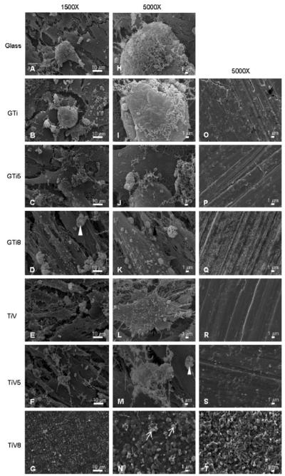
Fig. 5.
hFOB 1.19 adhesion after 14-day culture as assessed by SEM on glass coverslips (a, h), GTi (b, i), GTi5 (c, j), GTi8 (d, k), TiV (e, l), TiV5 (f, m), and TiV8 (g, n) disks. Most samples exhibited osteoblasts with cell projections (black arrow), fibrous networks that may correspond to collagen (black arrowhead) and sponge-like structures that may correspond to mineral nodules (white arrowheads). On TiV8 only were observed structures that resemble cell debris (white arrows). Negative control surfaces (incubated with culture media for 14 days but without cells) corresponding to each surface are displayed in (o)–(t) panels. Magnification: a–g = ×1,500, h–t = ×5,000. Scale bar: left = 10 μm, center and right = 1 μm

