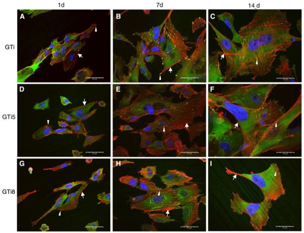
Fig. 6.

Visualization by confocal laser scanning microscopy of focal adhesions and cytoskeleton of hFOB 1.19 cells after 1 day (a, d, g), 7 days (b, e, h), and 14 days (c, f, i) of seeding on autoclaved (a–c), thermally oxidized at 500°C (d–f), and at 800°C (g–i) γ-TiAl surfaces. Cells exhibited stress fibers (arrows), focal contacts at the cell periphery and at the end of the stress fibers (arrowheads). Scale bar: 50 μm
