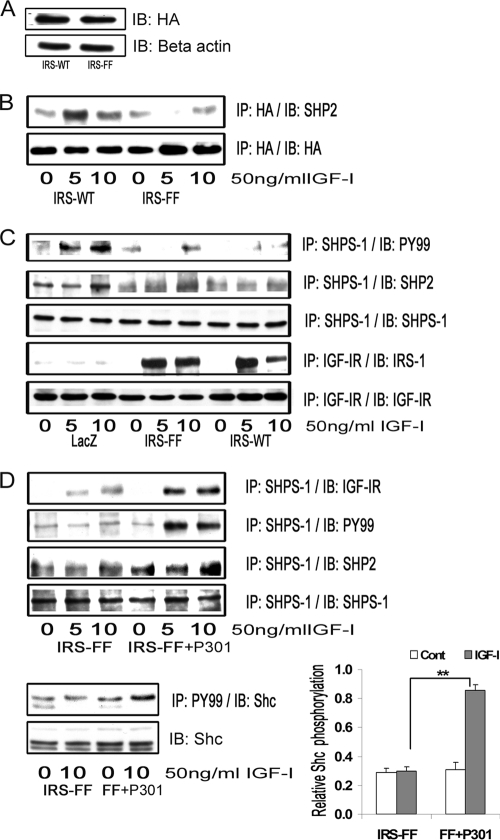
FIGURE 5.
IRS-1 sequesters SHP-2 and impairs its transfer to SHPS-1. A, VSMCs overexpressing IRS-WT and the IRS-FF mutant were serum-starved overnight and analyzed for recombinant protein expression. Cell lysates were immunoblotted (IB) for HA (top) and for β-actin (bottom). B, confluent cultures of IRS-WT and IRS-FF cells were serum-starved for 16 h and then exposed to IGF-I for the times indicated. Cell lysates were immunoprecipitated (IP) with anti-HA and immunoblotted for SHP-2 (top). The membranes were stripped and probed with anti-HA antibody (bottom). C, confluent cultures of control LacZ cells, IRS-WT cells, and IRS-FF cells were serum-starved overnight and stimulated with IGF-I for 5 or 10 min. The cell lysates were immunoprecipitated with anti-SHPS-1 and immunoblotted for Tyr(P)99 (top panel) or SHP-2 (second panel). They were stripped and reprobed with anti-SHPS-1 antibody (third panel). Lysates from similar experiments were immunoprecipitated with anti-IGF-IR and immunoblotted for IRS-1 (fourth panel). The blots were stripped and reprobed for IGF-IR (bottom panel). D, confluent IRS-FF cell cultures were serum-starved for 14–16 h and incubated with or without cell-permeable IGF-IR/IRS-1 peptide 301 (20 μg/ml) for 2 h followed by IGF-I for either 5 or 10 min. Cell lysates were immunoprecipitated with anti-SHPS-1 and immunoblotted for IGF-IR (first panel), Tyr(P)99 (second panel), SHP-2 (third panel), or SHPS-1 (fourth panel). Lysates were immunoprecipitated with Tyr(P)99 (PY99) and immunoblotted with anti-p52shc (fifth panel) or immunoblotted directly for total Shc (bottom panel). The bar graph shows the relative increase in Shc phosphorylation for at least three independent experiments. Error bars, mean ± S.E. **, p < 0.01 when increase in Shc phosphorylation in response to IGF-I is compared between IRS-FF mutant cells with or without peptide 301.

