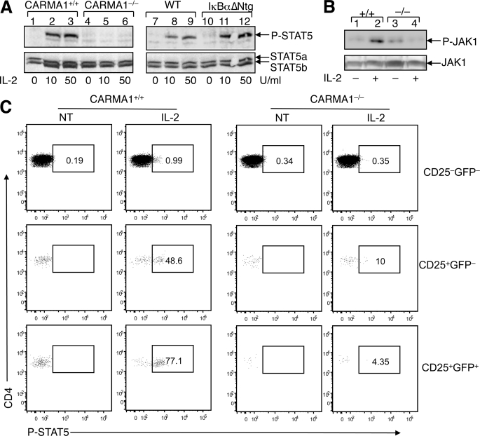
FIGURE 5.
CARMA1−/− nTreg precursors have IL-2 signaling defect. A, total thymocytes were isolated from CARMA1+/+ and CARMA1−/− mice or IκBαΔNtg and its control non-transgenic mice. The cells were stimulated with the indicated doses of IL-2 for 20 min and lysed for IB analysis of tyrosine-phosphorylated STAT5 (P-STAT5) (upper panel). The IB membrane was re-probed with regular anti-STAT5 to show the expression level of STAT5a and STAT5b (lower panel). B, total thymocytes from CARMA1+/+ (+/+) and CARMA1−/− (−/−) mice were incubated for 20 min either in the absence (−) or presence (+) of IL-2 (50 units/ml). JAK1 was isolated by IP (using anti-JAK1) followed by IB to detect phosphorylated JAK1 (P-JAK1) using anti-phospho-JAK1 (Tyr-1022/1023) (upper panel). Total JAK1 level in cell lysates was detected by IB using anti-JAK1 (lower panel). C, CARMA1+/+Foxp3-EGFP and CARMA1−/−Foxp3-EGFP thymocytes were stimulated with IL-2 (50 units/ml) for 30 min and immediately subjected to phospho-flow cytometry to detect tyrosine-phosphorylated STAT5 (P-STAT5) within the subsets of CD4+ single positive thymocyte populations. GFP was used as a marker for Foxp3 expression. Cells were gated on the CD25−GFP− cells, the CD25+GFP− Treg precursors, and the CD25+GFP+ mature Tregs. The percentage of P-STAT5-positive cells within each of the subsets is indicated.

