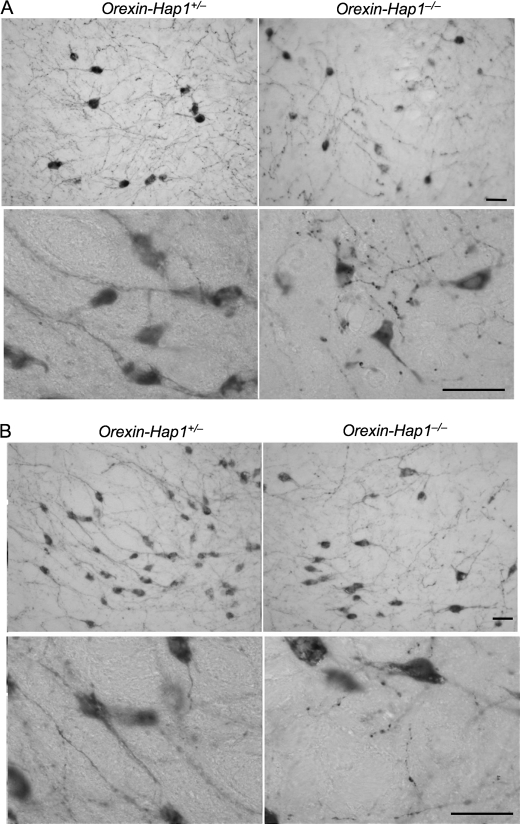
FIGURE 5.
Neuritic degeneration of orexin neurons in Orexin-Hap1−/− mice. Orexin-A immunohistochemical staining of the dorsomedial hypothalamic nucleus (A) and the lateral hypothalamic nucleus (B) of Orexin-Hap1+/− and Orexin-Hap1−/− mice. Low (×20, upper panels) and high (×65, lower panels) magnification images showing fragmented neurites of orexin neurons in Orexin-Hap1−/− mice. Scale bars, 10 μm.

