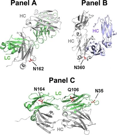
FIGURE 6.
Crystal structure of IgG2 Fab and Fc domains showing the location of non-consensus oligosaccharide structures. In Panel A, the IgG2 Fab crystal structure is rotated so that the HC Fd (gray) is in the foreground and the LC (green) is in the background and the non-consensus site at Asn-162 is shown in red. Panel B shows IgG2 Fc homodimer crystal structure with one chain colored gray and the other modified chain colored blue and the NCG site at Asn-360 shown in red. In Panel C, the IgG2 Fab crystal structure is oriented to show the occupied Asn and Gln residues (red) at positions 164, 106, and 35, respectively, from left, occurring on the LC (green).

