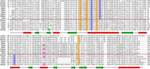
FIGURE 2.
Analysis of the primary structures of PrxQ proteins. Amino acid sequence alignments of various PrxQ from PrxQα and PrxQβ subfamilies. The red line separates PrxQ proteins into PrxQα (upper section) and Prxβ (lower section). Conserved peroxidatic and resolving cysteine residues are highlighted in blue. Conserved residues of the active site are highlighted in orange. XfPrxQ Cys-23 and Cys-101 are highlighted in green and magenta, respectively. PrxQα subfamily is represented by proteins as follows: ScDot5, S. cerevisiae DOT5 (GI: 731778); AtPrxQ, A. thaliana PrxQ (GI: 9279611); PoPrxQ, Poplar PrxQ (Populus balsamifera subsp. trichocarpa × Populus deltoides; GI: 42795441); SlPrxQ, Sedum lineare PrxQ (GI: 75336180); CdBcp, Clostridium difficile Bcp (GI: 126699430); EcBcp, E. coli Bcp (GI: 1788825); HiBcp, Haemophilus influenzae Bcp (GI: 1573220); KpBcp, K. pneumoniae Bcp (GI: 152971345); MtBcp, Mycobacterium tuberculosis Bcp (GI: 2791423); and StBcp, S. typhimurium Bcp (GI: 16765811). PrxQβ subfamily is represented by proteins as follows: RsPrxQ, Rhodobacter sphaeroides PrxQ (GI: 77387949); XcBcp, X. campestris Bcp (GI: 66768804); AvBcp, Anabaena variabilis Bcp (GI: 75906708); SyBcp, Synechocystis sp. (GI: 16329318); TeBcp, Thermosynechococcus elongatus Bcp (GI: 22298737); and XfPrxQ, Xylella fastidiosa PrxQ (GI: 9105889). The secondary structure of XfPrxQ C47S, obtained by PROCHECK (52), is shown below its sequence (green arrows representing β-sheets and red rectangles for α-helices).

