FIGURE 6.
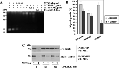
Gelatinolytically active MT6-MMP does not support cell migration. A, gelatin zymography of MT6F-PLC and MT6F-Nonidet P-40 (0.5–2.5 pmol), MT6CAT (0.05–2 pmol), and pro-MMP-2 (0.1–1 fmol). B, migration assay. HT-mock, HT-MT1, and HT-MT6F cells (1 × 10⁴) were allowed to migrate through type I collagen-coated Transwell inserts. Where indicated, GM6001 (50 μm) was added to the cells. The migration efficiency was calculated relative to HT-mock cells (100%). C, the uptake of MT6-MMP by cells. To prevent proteolysis of cellular MT1-MMP and MT6-MMP, HT and MCF7-MT6F cells (the top and bottom panels, respectively) were co-incubated with GM6001 (50 μm) for 16 h. The cells were then surface-biotinylated using membrane-impermeable, cleavable EZ-Link NHS-SS biotin and incubated for 30–60 min at 37 °C to stimulate the uptake of biotin-labeled plasma membrane proteins by the cells. Biotin-labeled protein was captured on streptavidin-beads, and the captured material was analyzed by Western blotting with the MT1-MMP and MT6-MMP antibodies. Where indicated, MESNA was used to release a biotin moiety from the cell surface-associated proteins. One representative experiment is shown. Multiple additional experiments generated similar results. IP, immunoprecipitation; WB, Western blot.

