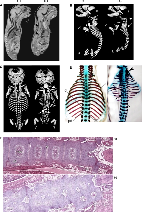
FIGURE 3.
A, micro-CT image of E18.5 non-transgenic control (CT) and COX-2 transgenic (TG) littermates in parasagittal orientation. Note the small thoracic cavity, collapsed lung (arrowhead), and loss of spinal curvature (arrow) in the transgenic fetus. B, lateral view of skeletal image analysis of E18.5 non-transgenic control and transgenic littermates. Ossified skeletal elements were visualized. Although the transgenic fetal skeletons exhibited reduced ossification in some of the dorsal cranial skull plates (arrowhead), which has a mesodermal origin, the rest of the skull, which is derived from neural crest, appeared normal. The long bones of the limbs were minimally affected except for the absence of deltoid tuberosity on the humerus in all transgenic fetuses (small arrow). The tympanic bulla appears to have been displaced caudally. In the transgenic fetus, normal vertebral curvature was lost (large arrow) due to malformation of vertebral components. C, dorsal view of skeletal image analysis of E18.5 non-transgenic control and transgenic littermates. In COX-2 transgenic fetuses, malformation occurred along the entire vertebral column and rib cage. Split ossification center (small arrow), fusion of pedicles with ossification center (large arrow), and fusion between pedicles of cervical vertebrae (arrowhead) were observed. Ribs were thin and discontinuous. D, dorsal view of Alcian blue/Alizarin red staining of E18.5 transgenic and control littermates. Cartilaginous elements are shown in blue, and ossified elements are shown in red. The forelimbs and shoulder girdles were removed for ease of viewing. Fusion of pedicles to ossification centers (small arrow) and dual ossification centers of vertebral bodies and fusion between ossification centers (large arrow) were observed. Fusions between pedicles of cervical vertebrae were displayed (arrowhead). The ribs were often thin and discontinuous, and the proximal end of the ribs did not contact the vertebral bodies. id, intervertebral disk; oc, ossification center; pd, pedicle; r, rib. E, hematoxylin/eosin staining of frontal section of E18.5 control and transgenic littermates (magnification, ×4). The right side is anterior. Note vertebral fusion and incomplete development of intervertebral disks and medullary cavity. me, medullary cavity; vb, vertebral body.

