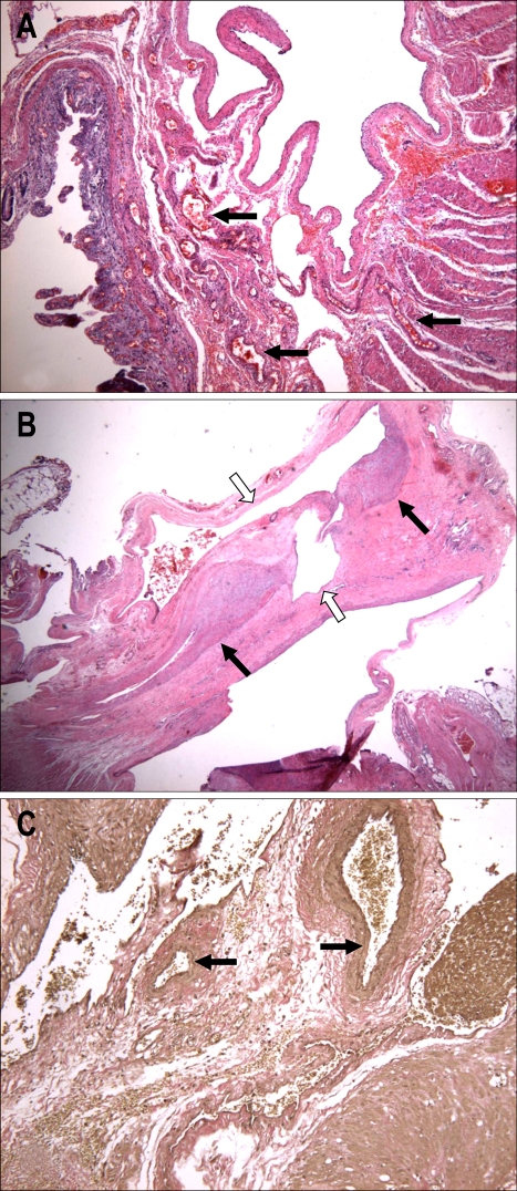
Fig. 4.
(A) Irregularly enlarged angiodysplastic vessels with rupture and hematoma formation in the submucosa. (B) Thick- and thin-walled vascular channels (black and white arrows, respectively) in the submucosa (hematoxylin & eosin stain; ×10). (C) Internal elastic fibers (black arrow) in thick artery-like vessels (elastin stain; ×100).

