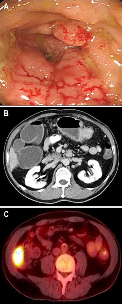
Fig. 1.
(A) Colonoscopy showed a bulky mass encircling the transverse colon and luminal narrowing. (B) CT showed a poorly enhancing mass in the proximal transverse colon with pericolic infiltration and regional lymph node enlargement. (C) Positron-emission tomography revealed abnormal fluorodeoxyglucose uptake in the cecum.

