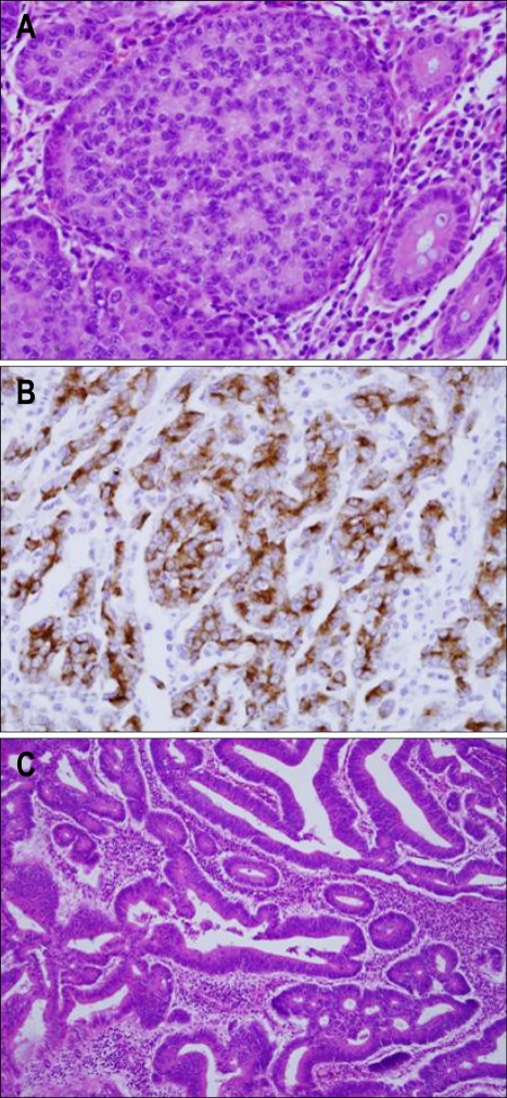
Fig. 3.
Histological and immunohistochemical examination. (A) Rosettes in the solid tumor nests and large polygonal cells (H&E stain, ×400). (B) Immunohistochemical staining revealed positive findings for chromogranin (Immunohistochemical stain for chromogranin, ×400). (C) Pathological examination of the mass in the cecum revealed an adenocarcinoma (H&E stain, ×100).

