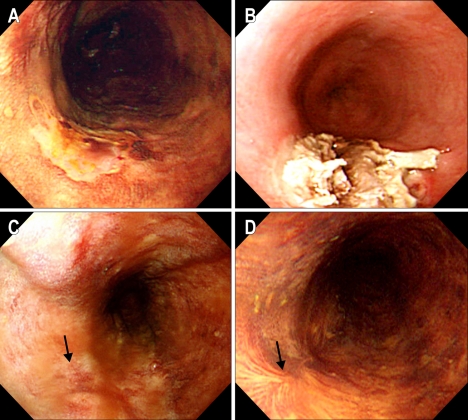
Fig. 1.
(Case #9) (A) Before PDT. A slightly depressed lesion unstained with Lugol's solution is seen on the mid-esophagus. (B) Two days after PDT. Endoscopy shows coagulation necrosis with ulcer at the PDT treated lesion. (C) One month after PDT. The previously PDT-induced ulcerative lesion have healed. (D) Five months after PDT. The scar well stained with Lugol's solution is seen at the previously cancerous lesion, and there is no remaining tumor in the biopsied specimens.

