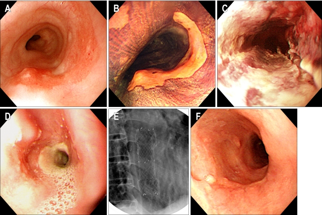
Fig. 2.
(Case #1) (A, B) A flat reddish lesion unstained with Lugol's solution is seen on the mid-esophagus (the biopsied shows specimen squamous cell carcinoma). (C) Two days after photodynamic therapy. Endoscopy shows circumferential coagulation necrosis with an ulcer at the PDT treated lesion. (D) Two months after PDT. Endoscopy shows luminal narrowing with fibrous scarring at the site of the PDT-treated lesion. (E) Fluoroscopic image shows a metal stent at the site of the esophageal stricture. (F) Endoscopy shows the improvement at the stricture site 2 months after stent removal.

