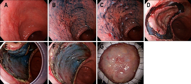
Fig. 4.
ESD procedures. (A) A depressed type EGC is noticed on the anterior wall of the antrum. (B) Indigo carmine dye is sprayed to detect the tumor boarder. (C) Markings are done by needle knife with coagulation current. (D) Mucosal cutting is done with IT knife using ENDO-CUT mode. (E) Dissecting submucosal layer is done by the aid of IT knife with ENDO CUT mode. Attachment cap is applied to stretch submucosal tissue. (F) A large ESD defect is noticed after complete one piece resection without perforation. (G) Flattened ESD specimen is fixed with thin needles on plate.

